Arthoscopy
What is Arthoscopy?
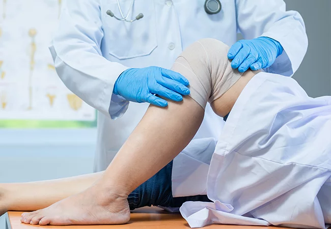
Arthroscopy is a surgical procedure that orthopaedic surgeons use to visualize and treat problems inside a joint.
Orthopaedic surgeons perform arthroscopic surgery by making a small incision in the patient’s skin and inserting pencil-size instruments that contain a small lens and lighting system to magnify and illuminate the structures inside the joint. A fibre optic cable transmits light to the end of the arthroscope inserted into the joint. An arthroscope is attached to a miniature camera so the surgeon can see inside the joint through this small incision rather than a larger one required for open surgery.
When is this recommended?
Inflammation.
For example, synovitis is a condition that causes the tissues surrounding the knee, shoulder, elbow, wrist, and ankle joint to become inflamed.
Acute or chronic injuries, including:
- Rotator cuff tendon tears
- Shoulder impingement
- Recurrent dislocation in the shoulder
- Meniscal (cartilage) tears in the knee
- Chondromalacia (wearing or injury of the cartilage cushion in the knee)
- Anterior cruciate ligament (ACL) tears with instability in the knee
- Carpal tunnel syndrome in the wrist
- Loose bodies of bone and/or cartilage, particularly in the knee, shoulder, elbow, ankle, or wrist
What is the Procedure?
A small incision (about the size of a buttonhole) will be made to insert the arthroscope. Several other incisions may be made to see other parts of the joint or insert other instruments.
Corrective surgery is performed with specially designed instruments that are inserted into the joint through accessory incisions. Originally, arthroscopy was simply a diagnostic tool used for planning standard open surgery.
After arthroscopic surgery, the small incisions will be covered with a dressing. You will be moved from the operating room to a recovery room. Many patients need little or no pain medication. Before being discharged, you will be given instructions on caring for your incisions, what activities to avoid, and which exercises you should do to aid your recovery.
What are the advantages?
Despite its widespread use in treating well-known athletes, arthroscopic surgery is an excellent tool for all orthopaedic patients, being less invasive than open surgery and generally more comfortable. Arthroscopy is usually performed as an outpatient procedure, and patients return home within a few hours after the procedure.